
Ultrasounds in Cleveland can detect pregnancies, fibroid tumors and ovarian or pelvic cysts/masses amongst a variety of other conditions.


Why Get Ultrasound Testing?
Ultrasounds provide noninvasive, three-dimensional (3D) imaging of a woman’s internal structures. A 3D ultrasound serves a multitude of functions, including identifying causes of pelvic pain, evaluation of menstrual issues, detection of ovarian cysts or uterine fibroids, and pregnancy monitoring, in addition to various other applications.
How It Works
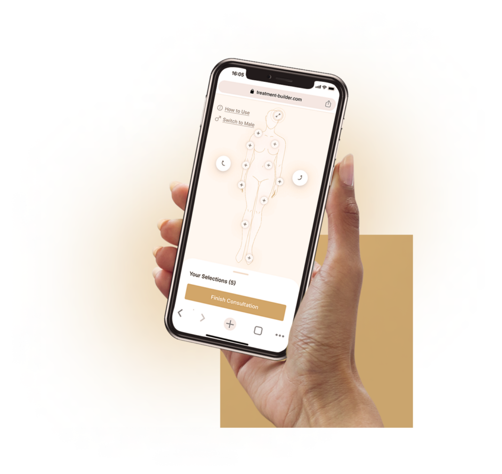
Step 1: Consultation
Before you begin any of our treatments, you will first talk with one of our providers for an initial consultation. They will speak with you about what issues you are experiencing and your medical history, then conduct diagnostic tests, which may include an ultrasound. This can be done during your consultation or during a separate appointment.
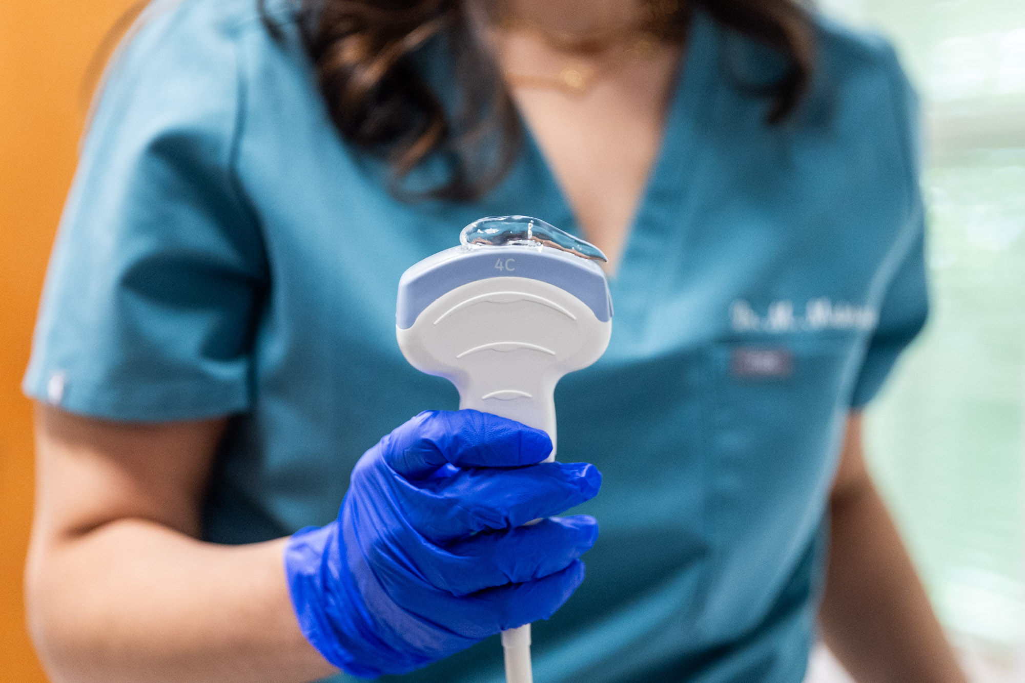
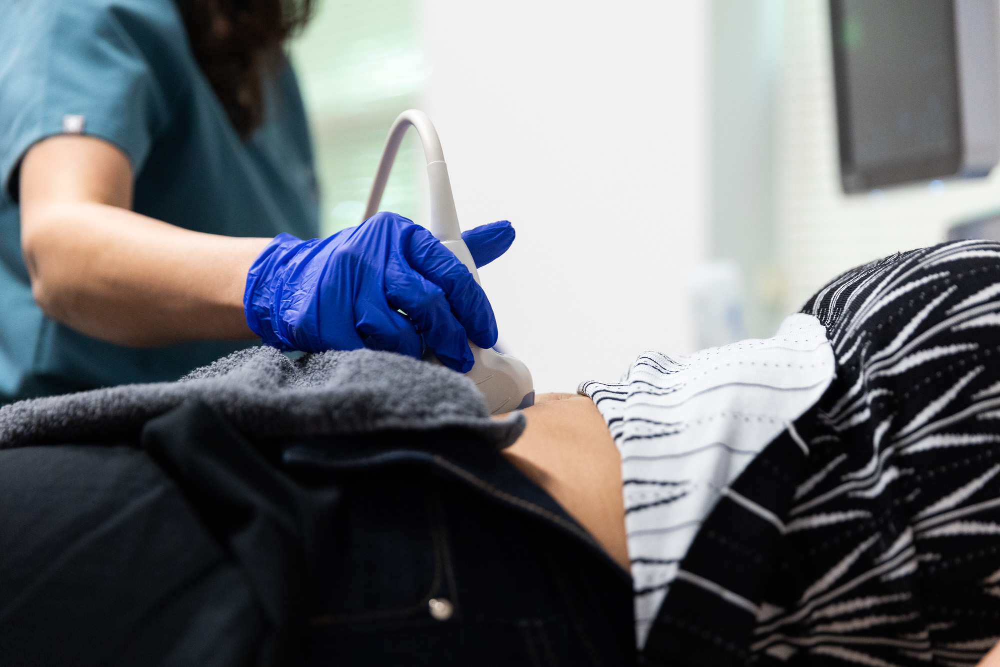
Step 2: Testing
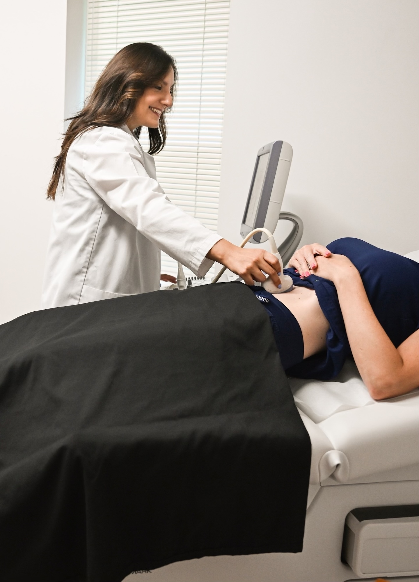
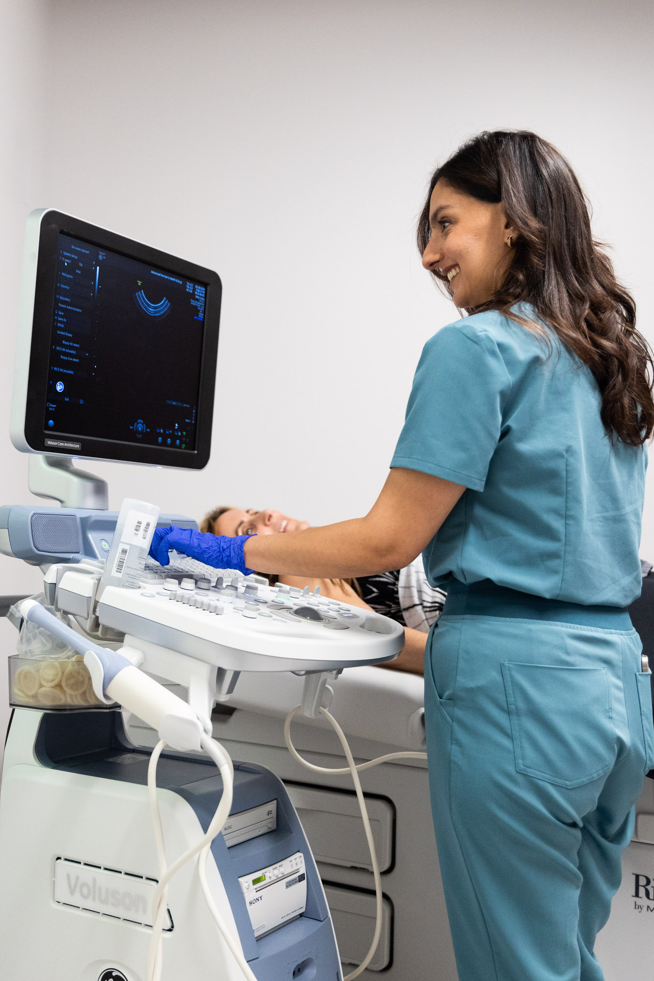
Your appointment may include either an abdominal or transvaginal ultrasound, or both. For abdominal ultrasounds, a gel will be applied to your abdomen, and then a wand will be used to run across your skin to get imaging of your abdomen. Transvaginal ultrasounds use a wand that is inserted into the vaginal canal to capture images of your pelvic structures.
Step 3: Follow-Ups
Once you are finished with your ultrasound in Cleveland, your provider will talk with you about your results. These results will help determine what medical conditions you have and help you and your provider determine the right treatments to help.
Our Benefits
Our team is woman-led and woman-owned, meaning we understand how important it is that your issues are resolved. At Advanced Women’s Health, you find even more benefits than with other women’s health clinics.
Comfort
The health issues we talk about with patients can be very sensitive, which can understandably make patients feel uncomfortable. We are dedicated to making sure you feel safe and supported when walking into our building and talking with our staff, ensuring you get the high-quality care you deserve.
Choices
While we are two separate businesses, we work together in the same building to achieve the same goal: helping women overcome their health and wellness issues. No matter if you need gynecological, wellness, or aesthetics services, our treatments will help you feel comfortable and confident in your body.
Clarity
Ensuring you have the best care possible is our #1 goal. We do that by making sure we have a clear picture of what your concerns are and asking plenty of questions that will help us determine the best possible care options for you. On the other hand, we also answer all the questions you have, ensuring you know every step that will be taken for your care.
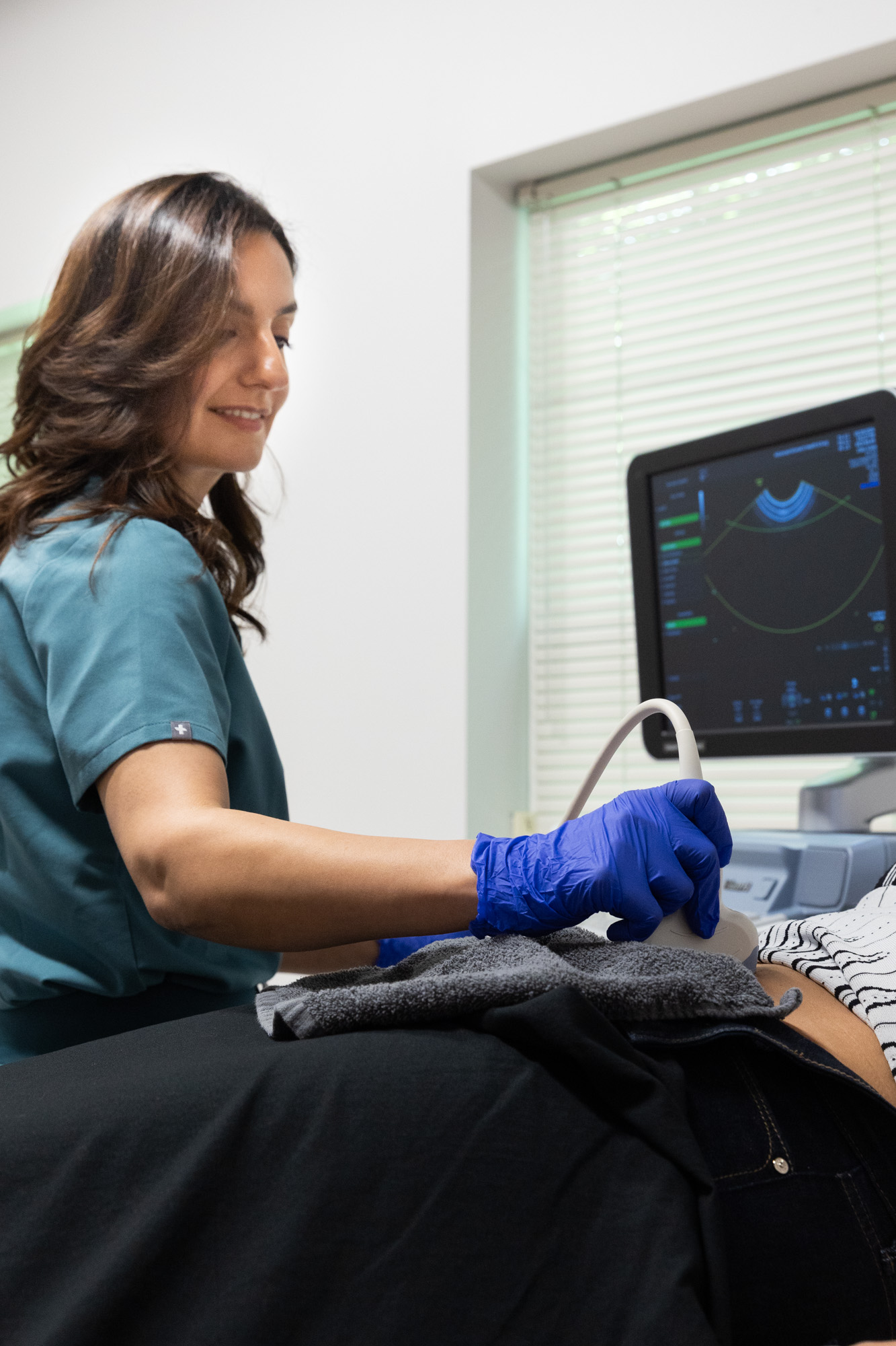
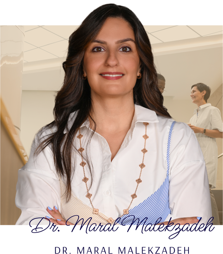
Meet Our Providers

Our exceptional team delivers high-quality care to our patients, including tests. With our experts by your side, we can quickly diagnose and treat whatever issues you may be facing. See our team page and meet the incredible staff who will be a part of your care team.
Related Services
Request an Appointment
Ready to take the first step towards feeling better in your body? Schedule a consultation today, and find your issues with an ultrasound in Cleveland.


Working Together for Women’s Health
The Well Westlake and Advanced Women’s Health & Surgery may be independent practices, but they share a common building for a common goal: making women’s lives better.
Each practice works synergistically with the other to provide the best holistic array of women’s health choices. Established by physicians who are also family (mother and daughters), and staffed with other skilled female physicians, our conjoined practices deliver the care you need—head to toe, inside and out.
Frequently Asked Questions
Your provider may suggest you have a full bladder before your ultrasound in Cleveland, but otherwise, there is no preparation you need to do before.
You will need to wear clothing that allows us access to your abdomen and pelvis, otherwise you may have to change into gowns provided.